What is an MEA (multielectrode array or microelectrode array)?
Microelectrode arrays – also known as “multielectrode arrays” or MEAs – are powerful tools used in neuroscience, cardiac research, and pharmacology to study the electrical activity of excitable tissues like neurons and cardiac cells. But what exactly is an MEA system, and how do they work?
Microelectrode arrays (also known as “multielectrode arrays” or MEAs) are powerful tools used in neuroscience research, cardiac research, pharmacology, and more to study the electrical activity of cells, particularly excitable tissues like neurons and cardiac cells. MEAs consist of an array of tiny electrodes that can simultaneously stimulate and record electrical signals from multiple cells or tissue regions. This allows researchers to capture detailed single-unit and field potential data.
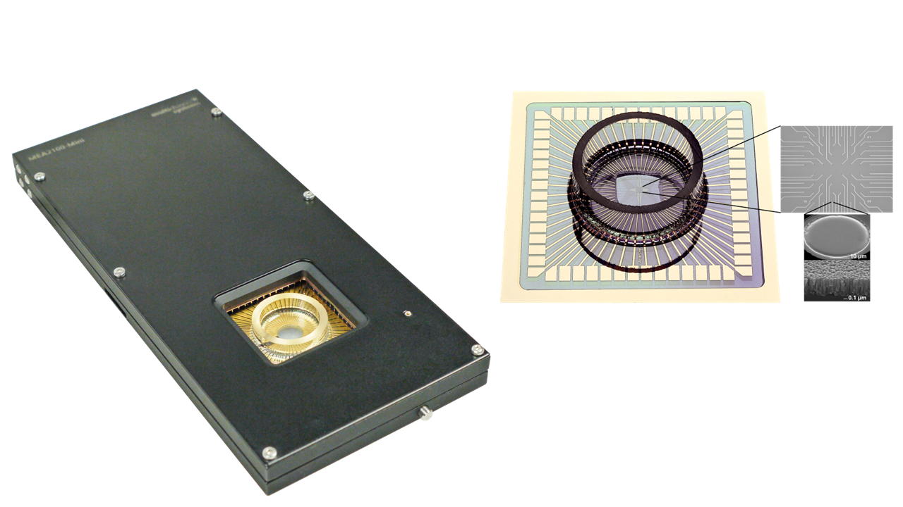
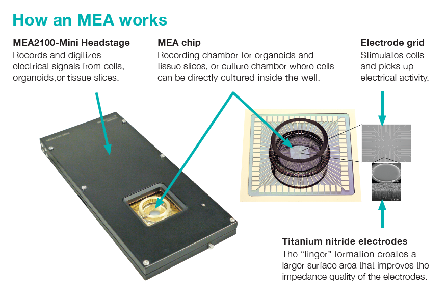
How an MEA Works
An MEA functions by placing an electrode grid in close contact with the tissue or cells being studied. These electrodes are capable of picking up spontaneous electrical signals as well as those evoked through chemical or electrical stimulation. The array structure allows researchers to stimulate cells from any specific electrode and observe how nearby cells respond. This provides valuable insights into cellular communication and behavior.
The components of a typical MEA system include:
- MEA chip: The recording chamber where organoids, tissue slices, or cultured cells reside during the experiment.
- Electrode grid: This grid stimulates cells and picks up their electrical signals.
- Titanium nitride electrodes: The unique "finger" design of these electrodes increases surface area, improving signal quality.
Why Use an MEA?
- Non-invasive: Unlike other techniques, MEAs do not damage cells, preserving their natural behavior.
- Easy Setup: MEAs are simpler and quicker to set up compared to more complex methods like patch clamping.
- High Throughput: Researchers can simultaneously record from dozens or even hundreds of locations, capturing a broad array of data in a single experiment.
- Real-time Monitoring: MEAs allow for continuous, long-term observation of cellular behavior, providing a dynamic look into how cells function over time.
- Versatile Integration: MEA systems can easily be integrated with other technologies, such as optical imaging, enhancing their research capabilities.
MEA Applications
MEAs are used in various fields of research and development, including:
- Neuroscience: To study neural communication and network behavior, as well as to investigate abnormal neural activity. MEAs can be used in organoid electrophysiology.
- Cardiac Research: Assess the electrical properties of cardiac cells and test new drugs for their effects on the heart.
- Pharmacology & Drug Testing: MEAs are crucial for drug screening, helping researchers observe how compounds affect cellular activity, especially in neurons and cardiac cells.
- Safety Pharmacology & Toxicology: Evaluate potential drug-induced issues such as hERG channel blockade and QT prolongation.
- Brain/Machine Interfaces: MEAs facilitate the development of robotic systems that interact with living tissues, like robotic arms controlled by neural activity, offering potential applications in prosthetics.
MEAs represent a significant advancement in the ability to study and monitor the electrical behavior of cells in real-time. Whether used in neuroscience, cardiac research, or drug development, their non-invasive nature and high throughput make them a go-to tool for modern research. With ongoing innovations in MEA technology, the possibilities for discovery continue to expand, offering new ways to understand complex cellular interactions and develop therapeutic solutions.