Exploring Retinal Development by Using Retinal Organoids and MEA Technology
Through our collaboration with Multi Channel Systems, the Ophthalmology Department at Radboudumc in the Netherlands is utilizing Mesh MEAs to capture dynamic retinal activity.
Cutting-edge advancements in stem cell–derived organoids and organ-on-chip technologies are transforming retinal research. Retinal organoids, developed from human induced pluripotent stem cells (hiPSCs) of both healthy and patient donors, offer promising in vitro models which closely mimic the human eye. These systems provide ethical alternatives to animal testing and open doors to understand mechanism of retinal diseases and personalized therapies.
However, current models still face challenges, including limited neuronal connectivity, lack of vascular structures, and inconsistent reproducibility. Encouragingly, breakthroughs in materials science and electronics help to bridge these gaps by enhancing tissue maturation and enabling real-time monitoring of neuronal activity.
Retinal organoids in low attachment 6-well plates
We developed mouse retinal organoids in low-attachment plates (i.e., floating in the well having flat surfaces), using timed chemical induction and growth factors to initiate early retinal differentiation. This approach enables early-stage retinal development like having retinal cell type including photoreceptors. However, retinal ganglion cells (RGCs) present majorly in the inner site of the retinal organoids, making them inaccessible to conventional 2D electrode recordings. This highlights a critical need for advanced platforms which enable both structural preservation and functional readouts.

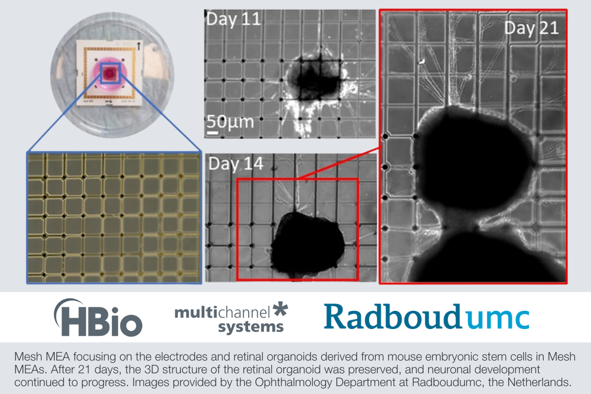
Retinal organoids derived from mouse embryonic stem cells at different developmental stages.
Preserving Structure, Capturing Function: Mesh Micro electrode arrays (MEAs)
Unlike conventional 2D electrodes, Mesh MEAs from Multi Channel Systems integrate directly within the 3D structure of the organoid, enabling stable and long-term access to inner retinal layers. By preserving neuronal architecture and capturing functional signals, Mesh MEAs open the door to more accurate light responsiveness assays and deeper insights into retinal development and disease.
Mesh MEAs are made of a flexible polyimide mesh with 60 electrodes positioned at the mesh junctions. When retinal organoids are plated to the mesh, they grow and develop around the mesh. This setup enables the electrodes to record from RGCs, located in the inner site of the organoid, and provides RGCs the opportunity to extend their protrusions outward. This innovative technique enhances the measurement of spontaneous activity, network connectivity, and responses to light stimuli.

Mesh MEA focusing on the electrodes and retinal organoids derived from mouse embryonic stem cells in Mesh MEAs. After 21 days, the 3D structure of the retinal organoid was preserved, and neuronal development continued to progress.

Electrical signals measured by Mesh MEA, capturing spontaneous activity (Top 1 and 2) as well as responses under light stimulation (Bottom). These measurements highlight the organoid’s ability to generate spontaneous neural activity and its responsiveness to external stimuli, offering insights into the functional properties of retinal ganglion cells.
Conclusion
We are laying the groundwork for developing functional human retinal organoids, unlocking new possibilities in disease modeling, personalized therapies, and next-generation vision research. Through our collaboration with Multi Channel Systems, we are utilizing Mesh MEAs to capture dynamic retinal activity. This breakthrough would not be possible without a multidisciplinary approach, where expertise in stem cell biology, materials science, and electronics come together to overcome complex challenges. By combining these fields, we are driving advancements in retinal diagnostics and therapies, pushing the boundaries of retinal research.
Article contributed by Zohreh Hosseinzadeh, PD Dr. and Yagmur Demircan Yalcin, PhD of the Ophthalmology Department at Radboudumc in the Netherlands.
To learn more about Mesh MEA, visit harvardbioscience.com/meshmea or contact our brand Multi Channel Systems online.