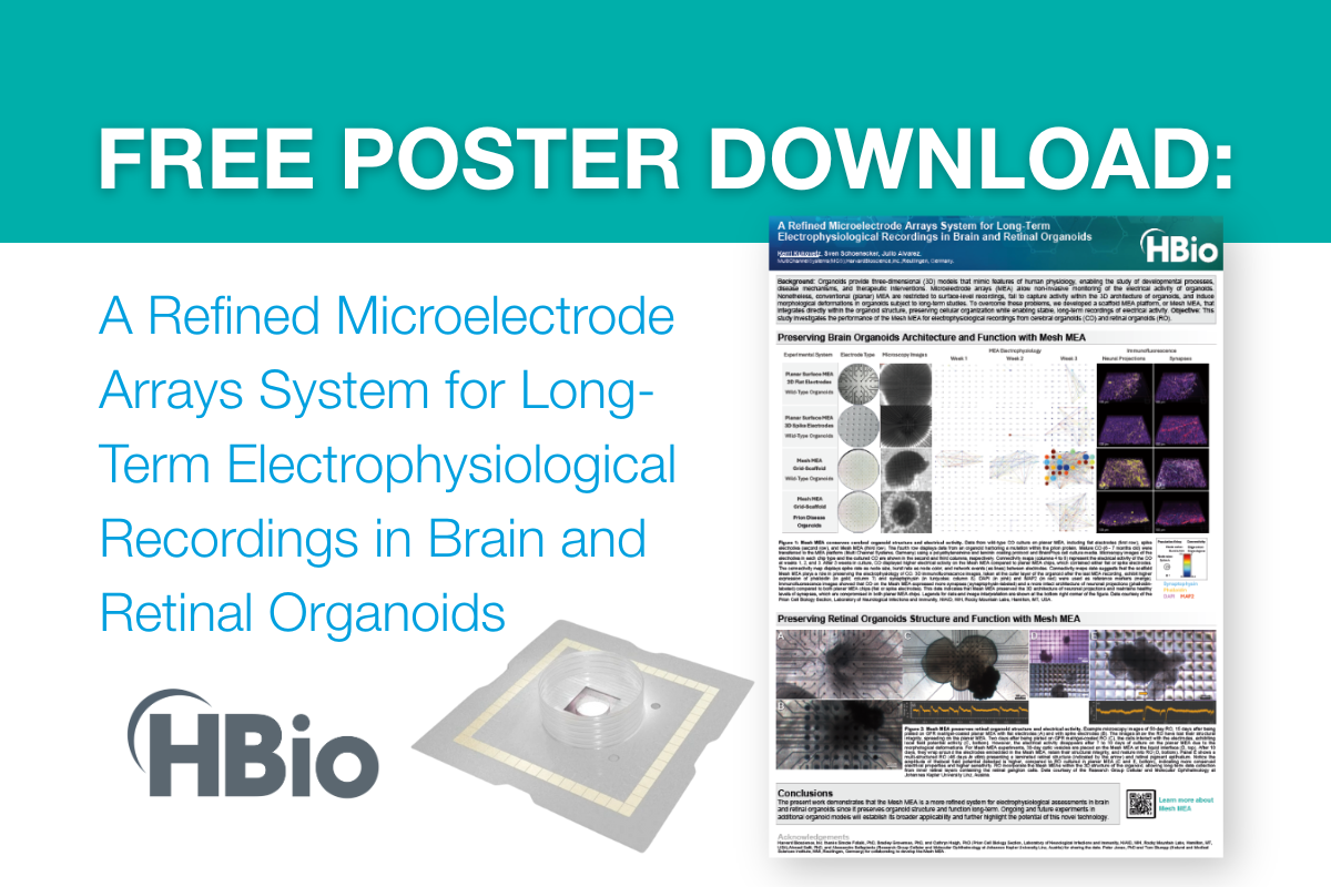
Using Organoids and MEA Technology to Research Retinitis Pigmentosa and Retinal Dystrophy
Retinitis pigmentosa (RP) and retinal dystrophy are debilitating genetic disorders that lead to progressive vision loss due to the degeneration of photoreceptor cells in the retina. Despite advances in genetic research, effective treatments remain limited, making the development of innovative research models crucial. Organoids and Mesh MEA (microelectrode array, also referred to as multielectrode array) technology from Multi Channel Systems have emerged as powerful tools in studying these diseases, providing insights into retinal function and potential therapeutic approaches.
Retinitis pigmentosa (RP) and retinal dystrophy are debilitating genetic disorders that lead to progressive vision loss due to the degeneration of photoreceptor cells in the retina. Despite advances in genetic research, effective treatments remain limited, making the development of innovative research models crucial. Organoids and Mesh MEA (microelectrode array, also referred to as multielectrode array) technology from Multi Channel Systems have emerged as powerful tools in studying these diseases, providing insights into retinal function and potential therapeutic approaches.
Organoids: A Breakthrough in Retinal Disease Modeling
Retinal organoids, derived from human pluripotent stem cells (hPSCs), offer a three-dimensional, physiologically relevant model that mimics the cellular architecture and function of the human retina. These self-organizing structures develop key retinal cell types, including photoreceptors, retinal ganglion cells (RGCs), and bipolar cells, making them ideal for studying the mechanisms underlying RP and other forms of retinal dystrophy.
By using patient-derived induced pluripotent stem cells (iPSCs), researchers can create patient-derived retinal organoids that reflect the genetic mutations responsible for RP. This approach allows for the study of disease progression, identification of early biomarkers, and testing of gene-editing or pharmacological interventions in a patient-specific manner.
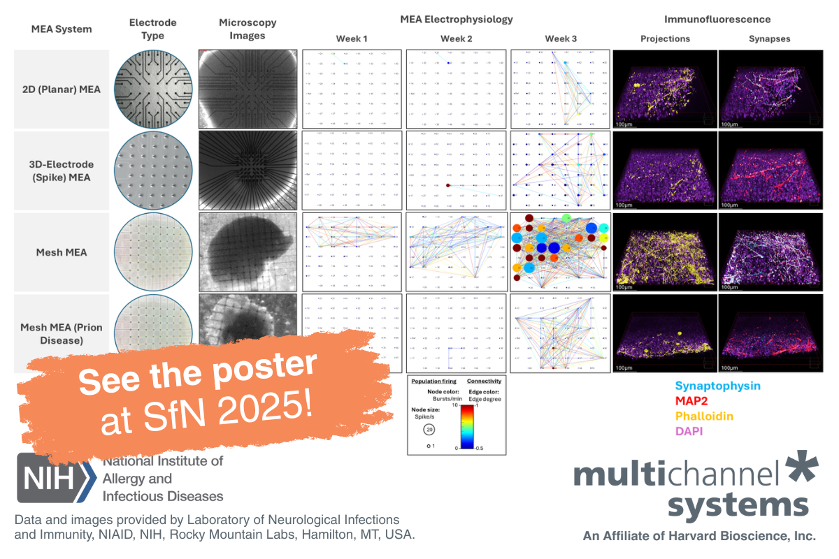
Mesh Microelectrode Arrays (MEA): A Window into Retinal Ganglion Cell Function
While retinal organoids replicate the structure of the retina, assessing their functional activity is essential for understanding disease pathology and evaluating treatments. RGCs are the most electrically active cells in the retina, and are centrally located within the retinal organoid, making it more challenging to record from these cells using traditional MEAs. Mesh MEA technology from Multi Channel Systems provides a solution to this problem and is an innovative method for measuring the electrophysiological activity of retinal cells.
Mesh MEAs consist of a flexible polyimide mesh with 60 electrodes located at the junctions of the mesh. Once retinal organoids are placed on the mesh, cells migrate through the mesh allowing the organoid to grow and develop around the mesh, thereby allowing the electrodes to record from the RGCs in the center of the organoid. Spontaneous activity, network connectivity, and responses to light stimuli can be more effectively measured with this new technique.
By integrating Mesh MEA with retinal organoids, researchers can:
- Characterize disease-specific functional deficits in patient-derived organoids.
- Test pharmacological compounds to assess their effects on retinal activity.
- Evaluate gene therapies by monitoring electrophysiological improvements after targeted interventions.
Advancing Therapeutic Strategies
The combination of organoid technology and Mesh MEA recordings provides a robust platform for drug discovery and precision medicine. For example, researchers from the Research Group Cellular and Molecular Ophthalmology at the Johannes Kepler University in Linz have used our Mesh MEAs to characterize the effects of a Retinitis Pigmentosa related gene (PRPF31) on the development and survival of RGCs. Preliminary data suggest that retinal organoids have improved survival, allowing for the recording of RGC activity over many days or weeks.

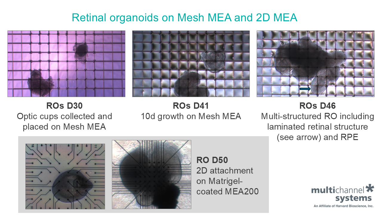
Day 50 retinal organoids 15 days after plating on GFR Matrigel-coated MEA200 30iR and 3D MEA lose their structural integrity, spreading on the MEA. Images courtesy of Johannes Kepler University Linz.

Day 50 retinal organoids two days after plating on GFR Matrigel-coated MEA200 30iR show interaction with the electrodes, presenting single spike activity and local field potential. Images courtesy of Johannes Kepler University Linz.

Day 30 optic vesicles manually collected and placed on Mesh MEA for 10 days retain their structural integrity and mature into stage 1 retinal organoids, compared to 10 days of culturing of D50 retinal organoids on GFR Matrigel-coated 2D MEA. Images courtesy of Johannes Kepler University Linz.

Retinal organoid on Mesh MEA presenting spikes and field potentials. Images courtesy of Johannes Kepler University Linz.
An improved understanding of how PRPF31 and other genetic variants play a role in the development of Retinitis Pigmentosa can eventually lead to the creation of more effective treatments and earlier detection of this disease.
As advancements continue, the synergy between organoids and MEA technology is expected to accelerate the development of targeted therapies for RP and retinal dystrophy, bringing us closer to potential cures for these currently incurable conditions.
Conclusion
The integration of retinal organoids and Mesh MEA technology represents an innovative and transformative approach to understanding and treating retinal degenerative diseases. By enabling patient-specific modeling, functional analysis, and drug screening, these tools are paving the way for innovative therapeutic strategies that could restore vision for individuals affected by RP and other retinal dystrophies.
Learn more about organoid electrophysiology and Mesh MEA.