Understanding Lung Fibrosis and the Future of Noninvasive Research Techniques
Lung fibrosis is a serious condition that limits lung function, and traditional research methods often rely on invasive techniques. However, Whole Body Plethysmography (WBP) offers a noninvasive solution for tracking fibrosis progression over time. This method allows researchers to monitor lung function in animal models without terminal procedures, providing valuable insights for developing effective treatments and understanding disease development.
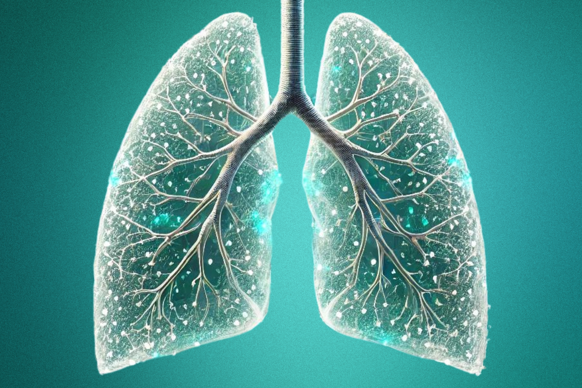
Lung fibrosis is a progressive and often devastating condition that impacts the lungs' ability to function properly. This disease occurs when lung tissue becomes damaged and scarred, leading to reduced lung compliance and impaired breathing. While the causes of lung fibrosis can vary—from environmental exposures and autoimmune diseases to infections—the condition is often classified as idiopathic pulmonary fibrosis (IPF) when no specific cause can be identified. In these cases, the development of the disease is particularly difficult to predict, making effective treatments elusive.
The Role of Plethysmography in Preclinical Fibrosis Research
In preclinical research, plethysmography has long been a cornerstone in assessing lung function. This method allows researchers to calculate important pulmonary endpoints, helping them understand how fibrosis affects lung compliance. However, traditional plethysmography comes with significant drawbacks. It’s an invasive process requiring anesthesia, tracheostomy, and mechanical ventilation. For rodent models, this is often terminal, meaning researchers can only gather data at a single point in time—typically the end of the study.
The challenge here is clear: researchers need a way to study fibrosis development longitudinally in the same subjects over time, across days, weeks, or even months. This continuous monitoring is vital for tracking both the progression of the disease and the effectiveness of potential treatments.
Is There a Noninvasive, Longitudinal Solution?
The good news is yes—Whole Body Plethysmography (WBP) offers a noninvasive alternative. Unlike invasive methods, WBP allows researchers to monitor unrestrained animals within a sealed chamber, measuring pressure fluctuations to derive respiratory flow and volume parameters. This technique has been trusted for decades in areas like asthma, opioid studies, infectious diseases, and sleep apnea. While WBP doesn’t provide direct measures of lung compliance, many researchers have discovered parameters within WBP data that strongly correlate with fibrosis development.
For example, researchers at Monash University tracked key parameters like Enhanced Pause (PenH), Inspiratory Flow (Tidal Volume / Inspiration Time), Inspiratory Duty Cycle (Ti/(Ti+Te)), and the Inspiratory-Expiratory Time Ratio (Ti/Te) over a 28-day study. These markers were shown to closely align with fibrosis progression. Similarly, other studies using bleomycin-induced fibrosis models have demonstrated that WBP can reliably detect impaired lung function as early as seven days after treatment. Enhanced Pause was found to predict terminal lung function deficits with a high degree of accuracy, making it a resourceful tool for ongoing research.
Why This Matters for Fibrosis Research
Using Whole Body Plethysmography at various stages of disease progression enables researchers to monitor how lung function changes over time. This is particularly beneficial for developing animal models of fibrosis and evaluating the impact of antifibrotic drugs or other therapies. With WBP, researchers can track lung function longitudinally without relying on invasive procedures. This approach not only provides more comprehensive data throughout the course of a study but also spares animals from terminal procedures, offering more humane and ethically sound research methods.
In a world where understanding lung fibrosis is critical to developing life-saving treatments, WBP stands as a promising tool for noninvasive, longitudinal study. As more researchers adopt this technique, we may see significant advancements in how we study, treat, and ultimately manage this debilitating condition.
Let’s chat!
Are you looking for solutions to enhance your respiratory research? Contact us today, and our team will connect with you!


