Applications
Organoid Research
Our MEA technology is at the forefront of organoid electrophysiology.

3D cellular structures self-assembling around the electrode-containing mesh. Photos courtesy of Jones’ lab, Biomedical Micro and Nano Engineering, NMI Natural and Medical Sciences Institute at the University of Tübingen.
Organoids are tissue-engineered, stem cell-based in vitro models that emulate human organs. They represent a unique experimental framework for studying human physiology and disease. However, limitations in readout platforms act as barriers to evolving organoid research.
In this sense, classical microelectrode arrays (MEA) fail to record data from inside organoids and induce collapsing and flattening of their structures on the surface of the 2D chips. Consequently, the physiological responses and the validity of the data are compromised.
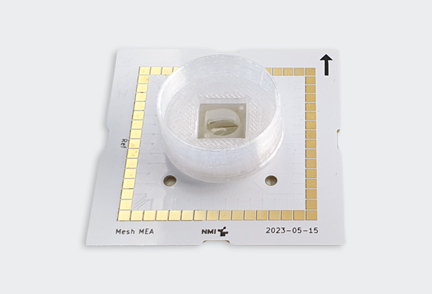
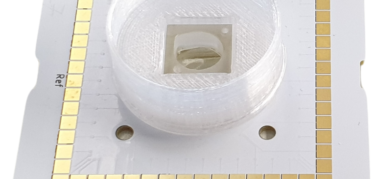
Our novel Mesh MEA platform mitigates these experimental challenges by preventing structural deformations and permitting the recording of electrical data from the inner sections of self-organizing 3D cultured organoids.
Key Benefits of Using Mesh MEA
- Record from inside an organoid: The cellular migration around the electrode-containing mesh enables monitoring signals from the organoid internal regions without compromising its structure.
- Collect electrophysiological data from an intact organoid: The mesh scaffold keeps the organoid suspended in a solution that protects and holds it in place and prevents morphological deformations of the organoid.
- Improve organoid maintenance: A perfusion system acts as a partial compensator for the lack of organoid vascularization.
- Long-term experiments: The undamaged organoid morphology and the longevity of maintenance allow multiple measurements in a single organoid.
- Flexible experimental settings: A user-defined air-liquid interface can be leveraged to facilitate organoid development and improve compound testing capabilities.

Figure 1: Neural activity of human brain organoids cultured in a 2D MEA2100 Mini platform (top panel) and in a 3D Mesh MEA platform (bottom panel). Raster plots show an increased neural spiking activity over time in organoids cultured in a Mesh MEA compared to classical MEA (MEA2100 Mini). Data courtesy of Hsieh’s lab, Department of Neuroscience, Developmental and Regenerative Biology at the University of Texas at San Antonio.
Applications
Performing electrophysiology research using organoids offers a cost-effective alternative to drug discovery and development. This innovative Mesh MEA technology is designed for emerging applications using organoids in research and discovery, precision medicine, safety pharmacology, and toxicology.
The system is suitable for all manner of 3D shapes, including:
- Cerebral, hippocampal, or midbrain organoids, neurospheres
- Cardiac organoids, spheroids, or cardiac bodies
- Pancreatic islets
- Retinal organoids
- 3D cell cultures and bioprinting organoids

Figure 2: Example images of the neuronal migration of human brain organoids on the Mesh MEA. Photos courtesy of Hsieh’s lab, Department of Neuroscience, Developmental and Regenerative Biology at the University of Texas at San Antonio.

Key Features of Mesh MEA
- The Mesh MEA features a 60-electrode flexible chip embedded in a slim 7 µm-thin polyimide mesh that promotes growth of cells around the electrodes
- Interelectrode distance of 200 µm and electrode (TiN) diameter of 30 µm
- Data is sampled at 50 kHz per channel at a 24-bit resolution
- Real-time signal detection and feedback
- Freely programmable digital signal processing
- Multiple inputs/outputs, including digital, analog, and audio
- SuperSpeed USB 3.0 ports
- Voltage and current stimulation possible
- Optical access from bottom
- Compatible with Multi Channel Systems' MEA2100 series

Though a Mesh MEA chip is ideal for organoid research, our brand Multi Channel systems also offers a range of 2D MEA options that will allow for the recording of cellular electrophysiological data – including CMOS, single-well, and multiwell options.
DOWNLOAD BROCHURE: OUR MEA TECHNOLOGIES
